MEDIA FOR THE ROUTINE GROWTH OF HUMAN CORNEAL CELLS
S.F.Webb
School of Biological Sciences,
University of East Anglia,
Norwich NR4 7TJ
UK
E-mail: s.f.webb@uea.ac.uk
Introduction
Materials
Methods
Results
Summary and conclusions
Human Corneal Cell cultures are successfully generated in our laboratories from un-transplantable corneo-scleral discs of all donor ages. Pure epithelial and fibroblast cells are grown routinely in EMEM supplemented with 20% FCS. Although effective for cell growth, the constituents in FCS are neither all identifiable nor quantifiable. As these cells will be used for miniature corneal constructs for growth and toxicity studies, it is preferable to use defined serum-free (SF) media where the constituents can be manipulated.
![]() THE PURPOSE OF THIS STUDY IS TO FIND SERUM-FREE
THE PURPOSE OF THIS STUDY IS TO FIND SERUM-FREE
MEDIA FOR THE ROUTINE GROWTH OF HUMAN CORNEAL CELLS
Tissues:
A. Observational Studies: Human Corneal SectionsSerum-free media tested (Clonetics):
All these media required several or all of the growth supplements listed
below:
Insulin, retinoic acid, hydrocortisone, transferrin, epinephrine, hEGF
epidermal growth factor, BPE (bovine pituitary extract) and T3 (tri-iodothyronine).
A. Observational Studies
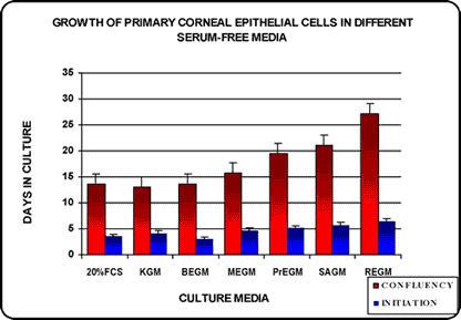
The criteria of culture success were the speed of culture initiation and the growth to confluency as compared with the control in FCS medium.
|
Fig. 1 |
Fig. 2 |
|
|
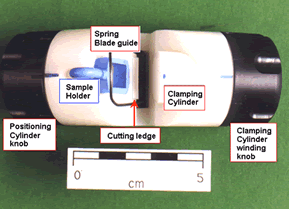
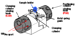
View of Microtome from the Top |
Microtome in cutting Position |
|
|
|
|
Fig. 3 |
|
Serial sections from Human Cornea |

|
B. Quantitative Studies
A) Observational Studies
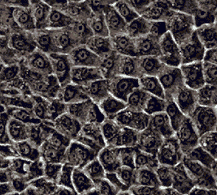
Epithelial Cultures (Fig 4)
-> MEGM -> SAGM = PrEGM ->
REGM Fig. 4
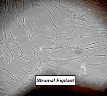
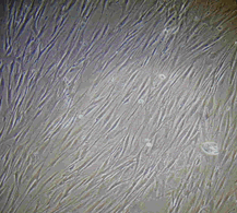
Stromal Fibroblast Cultures
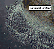
Epithelial Cultures (Click for an enlarged image):
|
|
|
2 Day-old culture |
10 Day-old culture |
Fibroblast Cultures (Click for an enlarged image):
|
|
|
7 Day-old culture |
11 Day-old culture |
B: Quantitative Studies on P=1 Cells
Fig. 5
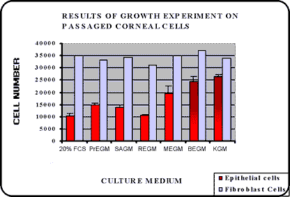
![]() At present KGM is the medium of choice as
BEGM requires
At present KGM is the medium of choice as
BEGM requires
more supplements as well as at twice the concentration.
Present work:
Human Corneal Constructs
2 methods of assembly are being investigated:
Acknowledgements:
Supported by The Humane Research Trust and the RSPCA.
Human corneas supplied by the Bristol Eye Bank.